| Identification | More | [Name]
D-Serine | [CAS]
312-84-5 | [Synonyms]
2-AMINO-3-HYDROXYPROPANOIC ACID
D-2-AMINO-3-HYDROXYPROPANOIC ACID
D-2-AMINO-3-HYDROXYPROPIONIC ACID
D-BETA-HYDROXYALANINE
D-(+)-SERINE
D-SERINE
H-D-SER-OH
(R)-2-AMINO-3-HYDROXYPROPIONIC ACID
β-Hydroxyalanine
D-SERINE 98.5%
D-Serine,>99%
D-2-Amino-3-Hydroxypropionic
(2R)-2-amino-3-hydroxy-propanoic acid
D-Serin
(R)-2-amino-3-hydroxypropanoic acid
D-SERINE extrapure
β-Hydroxyalanine, (R)-(-)-Serine, (R)-2-Amino-3-hydroxypropionic acid, 2-Amino-3-hydroxypropanoic acid, Ser
β-Hydroxyalanine, (R)-2-Amino-3-hydroxypropionic acid, Ser
Dserine1-norleucine4-valinamide25-β1-25-corticotrophin
DW75 | [EINECS(EC#)]
206-229-4 | [Molecular Formula]
C3H7NO3 | [MDL Number]
MFCD00004269 | [Molecular Weight]
105.09 | [MOL File]
312-84-5.mol |
| Chemical Properties | Back Directory | [Appearance]
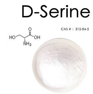
White powder | [Melting point ]
220 °C
| [alpha ]
-14.75 º (c=10 2 N HCl) | [Boiling point ]
197.09°C (rough estimate) | [density ]
1.3895 (rough estimate) | [refractive index ]
1.4368 (estimate) | [storage temp. ]
Store at RT. | [solubility ]
H2O: 0.1 g/mL, clear, colorless
| [form ]
Crystalline Powder | [pka]
2.16±0.10(Predicted) | [color ]
White | [Water Solubility ]
346 g/L (20 ºC) | [Merck ]
14,8460 | [BRN ]
1721403 | [InChIKey]
MTCFGRXMJLQNBG-UWTATZPHSA-N | [LogP]
-1.490 (est) | [CAS DataBase Reference]
312-84-5(CAS DataBase Reference) | [EPA Substance Registry System]
312-84-5(EPA Substance) |
| Safety Data | Back Directory | [Hazard Codes ]
Xi | [Risk Statements ]
R36/37/38:Irritating to eyes, respiratory system and skin . | [Safety Statements ]
S24/25:Avoid contact with skin and eyes .
S36:Wear suitable protective clothing .
S26:In case of contact with eyes, rinse immediately with plenty of water and seek medical advice . | [WGK Germany ]
3
| [RTECS ]
VT8200000 | [TSCA ]
Yes | [HS Code ]
29225000 |
| Questions And Answer | Back Directory | [Overview]
D-serine is the D-form of the amino acid serine, but is not used for the protein synthesis. Amino acids are among the most significant molecules in nature and exist in an l- and a d-form. The chemical and physical properties of l- and d-amino acids are enormously similar except for their optical characteristics[1]. During the emergence of life, only the l-amino acids were selected for the formation of polypeptides and proteins. Amino acids are not present in mammals and that d-amino acids were restricted to some bacteria and insects. Only a few decades ago, it was largely believed that free d- amino acids are not present in mammals and that d-amino acids were restricted to some bacteria and insects. Often, d-amino acids were called “unnatural” amino acids and they were considered to be the by-products of microorganisms metabolism.
The first report to show the presence of substantial quantities of free d-amino acids in mammalian tissues was by dunlop et al 1986 where, surprisingly, a large amount of d-aspartic acid[d- asp] in the cerebrum of a newborn rat and in the pituitary gland of an adult rat was reported[2]. A second d-amino acid, d-serine, was then identified in considerable amounts in the brains of rodents and man[3, 4]. Successive studies verified that some d- amino acids exist in the mammalian central nervous system(CNS) and peripheral tissues in, unpredictably, high concentrations that may exceed the level of l-amino acids occurrence[4]. The unanticipated detection of large amounts of endogenous d-serine in the brain, by hashimoto et al, initiated a series of studies from several laboratories that investigated the physiological role of endogenous d-serine. Recently endogenous d-serine has been associated with several physiological and pathological n-methyl-d-aspartate receptor(NMDAR)-reliant processes, including NMDAr transmission and synaptic plasticity[5-7], cell migration, and neurotoxicity[8-10].

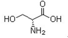
Figure 1 the chemical structure of D-serine | [Localization]
The distribution of d-serine is parallel to the distribution of nMda type glutamate receptors[4]. D-Serine has been detected at relatively high levels in certain areas in the adult brain with particularly high levels of nMdars, including cerebral cortex, hippocampus, thalamus, hypothalamus, amygdala, and retina. Nonetheless, brain regions, such as the hindbrain, pons, and medulla have nearly imperceptible levels of d-serine. Significantly, it has been demonstrated that d-serine is localized principally within glial cells[14, 5] in the retina, Stevens et al[4] reported the occurrence of d-serine in astrocytes and Mu?ller glia cells. Recently, several studies suggest that the synthesis, storage, and release of d-serine may not be limited exclusively to astrocytes, but rather may involve specific functions for certain cells[6].
| [Synthesis and Metabolism]
Humans can acquire D-serine through ingestion with food, derivation from gastrointestinal bacteria, liberation from metabolically stable proteins, which contain D-amino acids after racemization with ageing, and through biosynthesis from L-serine. Few data are available on the relative contributions of these four sources, but biosynthesis appears to be important. The enzyme serine racemase(SR) directly converts Lto D-serine in the presence of the co-factors pyridoxal 5-phosphate, magnesium and ATP[15-17]. SR also converts Dto L-serine, albeit with lower affinity[17]. D-Serine concentrations are thus highly related to L-serine concentration and thereby also to glycine concentrations[18]. Of the different pathways involved in L-serine biosynthesis, the glucose– 3-phosphoglycerate-3-phosphoserine–biosynthesis pathway is essential for normal embryonic development, especially for brain morphogenesis.[19] Consequently, D-serine concentrations in the developing CNS might also depend heavily on this pathway.
SR is highly expressed in the brain, with lower levels in the liver and small or no detectable expression in other tissues. In the brain, SR localizes to protoplasmic astrocytes in a pattern similar to D-serine.[16,17] Physiological synthesis of D-serine by SR in the glia was implicated by the strong spatiotemporal correlation between D-serine and SR[20] and by the decrease in D-serine concentrations in astrocytes after pharmacological inhibition of SR.[16] The cDNA encoding human SR has been cloned and D-serine synthesis by SR has been demonstrated in living cells after heterologous overexpression.[21] Whereas human serine hydratase does not contribute substantially to the degradation of L-serine to pyruvate, SR was found to catalyze, in addition to the racemase activity, the α,β-elimination of water from both L-serine and D-serine to form pyruvate and ammonia.[15,22] Under physiological conditions, pyruvate formation seems to equal or excess Dserine formation. Pyruvate formed by SR may be sufficient for the energy requirements of the astrocytes. This reaction further implies that SR is not only involved in D-serine synthesis, but also in D-serine metabolism as a mechanism to regulate intracellular Dserine levels.[22] Mammalian D-amino acids can be metabolized by the peroxisomal flavoprotein DAO, with the concomitant reduction of the co-factor flavin adenine dinucleotide(FAD). Physiological degradation of D-serine by DAO was suggested by the marked regional and developmental variation in DAO levels in a pattern reciprocal to D-serine levels.[20] Furthermore, Dao-/mice manifest an increase in D-serine levels, especially in areas with low levels in wild type animals such as the cerebellum and periphery[24]. The relatively unchanged D-serine levels in the forebrains of DAO-/mice imply that in these areas, other mechanisms might regulate D-serine concentrations[24,25].
| [Biological effects]
NMDAr neurotransmission
The evident association between the anatomical distribution of d-serine and the localization of the NMDAr suggests a functional relationship. NMDArs are largely distributed throughout the CNS and play a major role in glutamatergic synaptic transmission[26]. NMDArs are tetrameric ionotropic receptor channels that are major excitatory receptors in the brain; they play various roles in different physiological processes, such as nMda transmission, synaptic plasticity, and development[26].
Functional evidence for the contribution of endogenous D-serine to physiological nMdar co-activation was reported in a pioneer study by Mothet et al. in this study, addition of DAO, an enzyme that selectively degrades d-amino acids but not l-amino acids, to neural cell cultures resulted in depletion of endogenous d-serine and eventually noticeable reduction in nMdar activity[7]. This effect was fully reversed by the application of exogenous d-serine[7]. Subsequent studies demonstrated that endogenous d-serine is required for nMdar mediated lightevoked responses in the vertebrate retina[5, 11].
NMDArs play a major role in excitatory transmission and synaptic plasticity, such as long-term potentiation(LTP)[28]. D-Serine contribution to activity-induced synaptic plasticity was further confirmed when yang et al compared the ability to evoke LTP in cultured neurons between cells grown in direct contact with astrocytes and those grown without direct contact. Surprisingly, neurons that were not in direct contact with astrocytes failed to induce LTP. When the cells were supplemented with an exogenous source of d-serine, LTP was successfully induced[27]. Similarly, the contribution of d-serine to activity-induced synaptic plasticity in other brain areas, such as the hypothalamus, retina, and prefrontal cortex has been confirmed[12, 29, 30].
CNS development
The noticeably elevated d-serine concentrations in human and rodent CNS 4, 26 during the intense stage of embryonic and early postnatal CNS development provided the first evidence for a specific role for d-serine in CNS development. Supportive to this role, elevated d-serine concentrations coincide with transient expression[31] and increased activity[32-34] of nMdars. Likewise, Fuchs et al reported the presence of high d-serine concentrations in human cerebrospinal fluid(CSF) during the early postnatal period[35]. Moreover, excessive levels of d-serine have been detected in the cerebellum of neonatal rats, decreasing to very low levels in the third week of life as a result of the emergence of dao26. This temporary abundance of d-serine in the cerebellum corresponds with postnatal cerebellar development, in which granule cells migrate from the external to the internal granule cell layer in an nMdar-dependent manner[36]. Moreover, it has been shown that d-serine appears to be engaged in neuronal migration. DAO catalyzed degradation of d-serine and selective inhibition of Sr in eight day-old mouse cerebellar slices considerably suppressed granule cell migration, while d-serine appears to activate the migration through nMdar activation[36]. Supportive evidence for the d-serine role in migration is provided by the definite mass spectrometric identification of SR in the perireticular nucleus, a short-lived structure of the developing brain in humans proposed to be largely involved in neuronal migration[37].
Learning and memory
Long-term potentiation(LTP) of synaptic transmission in the hippocampus is broadly considered as one of the key cellular mechanisms underlying learning and memory in vertebrates96. It refers to an augmentation in signal transmission between neurons upon synchronic stimulation and is one of the fundamental processes of synaptic plasticity. D-Serine released from astrocytes and nMdar activation both appears to play a role in LTP induction. On the other hand, nMdar antagonists and enzymatic d-serine degradation suppressed LTP induction27. Further support provided from studies on SR knockout mice, where it had been shown that depletion of d-serine concentrations was directly related to an impaired nMdar transmission and attenuated ltp[38]. On the contrary, DAO knockout mice display high extracellular d-serine concentrations, improved NMDAr function, and enhanced hippocampal LTP[39, 40].
Studies assessing learning and memory decline occurring with aging revealed that SR expression, d-serine concentrations, nMdar-mediated synaptic potentials, and LTP were all drastically decreased in Ca1 hippocampal slices from aged rats when compared with young rats, and were all restored by exogenous d-serine[6, 41]. Similarly, hippocampal slices from a senescence-accelerated mouse model exhibited substantial and amplified LTP suppression with age, when compared to normal mice, which was overcome by D-serine supplementation. Collectively, these results strongly demonstrate the significance of d-serine for nMdar activation and subsequent LTP induction that underlies learning and memory.
| [Relation with diseases]
As it is involved in nMdar neurotransmission in the brain, nMdar-dependent plasticity, and developmental processes, it is not astonishing that dysregulation of d-serine signaling might also be involved in several pathologies, including neuropsychiatric and neurodegenerative diseases related to nMdar dysfunction. Intense stimulation of nMdars has been associated with considerable number of acute and chronic neurodegenerative conditions, including stroke, epilepsy, polyneuropathies, chronic pain, amyotrophic lateral sclerosis(ALS), Parkinson’s disease(PD), Alzheimer’s disease(AD), and Huntington’s disease(HD)[42].
| [References]
- lamzin vS, dauter Z, Wilson KS. how nature deals with stereoisomers. Curr opin Struct biol. 1995;5:830-6.
- dunlop dS, neidle a, Mchale d, dunlop dM, lajtha a. the presence of free d-aspartic acid in rodents and man. Biochem biophys res Commun. 1986;141:27-32.
- hashimoto a, nishikawa t, hayashi t, et al. the presence of free d-serine in rat brain. febS lett. 1992;269:33-6.
- hashimoto a, Kumashiro S, nishikawa t, et al. embryonic development and postnatal changes in free d-aspartate and dserine in the human prefrontal cortex. J neurochem. 1993;61: 348-51.
- gustafson eC, Stevens er, Wolosker h, Miller rf. endogenous dserine contributes to nMda receptor-mediated light-evoked responses in the vertebrate retina. J neurophysiol. 2007;98: 122-30.
- Junjaud g, rouaud e, turpin f, Mothet Jp, billard JM. age-related effects of the neuromodulator d-serine on neurotransmission and synaptic potentiation in the Ca1 hippocampal area of the rat. J neurochem. 2006;98:1159-66.
- Mothet Jp, parent at, Wolosker h, et al. d-Serine is an endogenous ligand for the glycine site of the n-methyl-d-aspartate receptor. PNAS. 2000;97:4926-31.
- Katsuki h, nonaka M, Shirakawa h, Kume t, akaike a. endogenous d-serine is involved in induction of neuronal death by n-methyl-d-aspartate and simulated ischemia in rat cerebrocortical slices. J pharmacol exp ther. 2004;311:836-44.
- Kartvelishvily e, Shleper M, balan l, dumin e, Wolosker h. neuron-derived d-serine release provides a novel means to activate n-methyl-d-aspartate receptors. J biol Chem. 2006; 281:14151-62.
- Katsuki h, Watanabe y, fujimoto S, Kume t, akaike a. Contribution of endogenous glycine and d-serine to excitotoxic and ischemic cell death in rat cerebrocortical slice cultures. Life Sci. 2007;81:740-9.
- Stevens er, esguerra M, Kim pM, et al. d-Serine and serine racemase are present in the vertebrate retina and contribute to the physiological activation of nMda receptors. PNAS. 2003;100:6789–94.
- Panatier a, theodosis dt, Mothet Jp, et al. glia derived d-serine controls nMda receptor activity and synaptic memory. Cell. 2006;125:775–84.
- Van horn Mr, Sild M, ruthazer eS. d-Serine as a gliotransmitter and its roles in brain development and disease. front Cell neurosci. 2013;7:39-52.
- Kreil g. peptides containing a d-amino acid from frogs and molluscs. J biol Chem. 1994;269:10967-70.
- Williams SM, Diaz CM, Macnab LT, Sullivan RK, Pow DV. Immunocytochemical analysis of D- serine distribution in the mammalian brain reveals novel anatomical compartmentalizations in glia and neurons. Glia 2006 March;53[4]:401-11.
- Kartvelishvily E, Shleper M, Balan L, Dumin E, Wolosker H. Neuron-derived D-serine release provides a novel means to activate N-methyl-D-aspartate receptors. J Biol Chem 2006 May 19;281[20]:14151-62.
- Yoshikawa M, Nakajima K, Takayasu N, Noda S, Sato Y, Kawaguchi M et al. Expression of the mRNA and protein of serine racemase in primary cultures of rat neurons. Eur J Pharmacol 2006 October 24;548[1-3]:74-6.
- Yasuda E, Ma N, Semba R. Immunohistochemical evidences for localization and production of D-serine in some neurons in the rat brain. Neurosci Lett 2001 February 16;299[1-2]:162-4.
- Bruckner H, Haasmann S, Friedrich A. Quantification of D-amino acids in human urine using GC-MS and HPLC. Amino Acids 1994;6[205]:211.
- Nagata Y, Masui R, Akino T. The presence of free D-serine, D-alanine and D-proline in human plasma. Experientia 1992 October 15;48[10]:986-8.
- Rotgans J, Wodarz R, Schoknecht W, Drysch K. The determination of amino-acid enantio- mers in human saliva with Chirasil-Val. Arch Oral Biol 1983;28[12]:1121-4.
- Stevens ER, Esguerra M, Kim PM, Newman EA, Snyder SH, Zahs KR et al. D-serine and serine racemase are present in the vertebrate retina and contribute to the physiological activation of NMDA receptors. Proc Natl Acad Sci U S A 2003 May 27;100[11]:6789-94.
- De Miranda J, Panizzutti R, Foltyn VN, Wolosker H. Cofactors of serine racemase that physi- ologically stimulate the synthesis of the N-methyl-D-aspartate[NMDA] receptor coagonist D-serine. Proc Natl Acad Sci U S A 2002 October 29;99[22]:14542-7.
- Wolosker H, Blackshaw S, Snyder SH. Serine racemase: a glial enzyme synthesizing D- serine to regulate glutamate-N-methyl-D-aspartate neurotransmission. Proc Natl Acad Sci U S A 1999 November 9;96[23]:13409-14.
- Wolosker H, Sheth KN, Takahashi M, Mothet JP, Brady RO, Jr., Ferris CD et al. Purification of serine racemase: biosynthesis of the neuromodulator D-serine. Proc Natl Acad Sci U S A 1999 January 19;96[2]:721-5.
- danysz W, parsons Cg. glycine and n-methyl-d-aspartate receptors: physiological significance and possible therapeutic applications. pharmacol rev. 1998;4:597-664.
- yang y, ge W, Chen y, et al. Contribution of astrocytes to hippocampal long-term potentiation through release of d-serine. proc natl acad Sci uSa. 2003;100:15194-9.
- Constantine-paton M, Cline ht, debski e. patterned activity, synaptic convergence, and the nMda receptor in developing visual pathways. annu rev neurosci. 1990;13:129-54.
- henneberger C, papouin t, oliet Sh, rusakov da. long-term potentiation depends on release of d-serine from astrocytes. nature. 2010;463:232-6.
- Stevens er, gustafson eC, Sullivan SJ, esguerra M, Miller rf. light-evoked nMda receptor-mediated currents are reduced by blocking d-serine synthesis in the salamander retina. neuroreport. 2010;21:239-44.
- Slater p, McConnell Se, d'Souza SW, barson aJ. postnatal changes in n-methyl-d-aspartate receptor binding and stimulation by glutamate and glycine of[3h]-MK-801 binding in human temporal cortex. br J pharmacol. 1993;108:1143-9.
- Crair MC, Malenka rC. a critical period for long-term potentiation at thalamocortical synapses. nature. 1995;375:325-8.
- feldman de, nicoll ra, Malenka rC, isaac Jt. long-term depression at thalamocortical synapses in developing rat somatosensory cortex. neuron. 1998;21:347-57.
- ramoa aS, McCormick da. enhanced activation of nMda receptor responses at the immature retinogeniculate synapse. J neurosci. 1994;14:2098-105.
- fuchs Sa, dorland l, de Sain-van der velden Mg, et al. d-Serine in the developing human central nervous system. ann neurol. 2006;60:476-80.
- Komuro h, rakic p. Modulation of neuronal migration by nMda receptors. Science. 1993;260:95-7.
- hepner f, pollak a, ulfig n, yae-Kyung M, lubec g. Mass spectrometrical analysis of human serine racemase in foetal brain. J neural transm. 2005;112:805-11.
- basu aC, tsai ge, Ma Cl, et al. targeted disruption of serine racemase affects glutamatergic neurotransmission and behavior. Mol psychiatry. 2009;14:719-27.
- almond Sl, fradley rl, armstrong eJ, et al. behavioral and biochemical characterization of a mutant mouse strain lacking d-amino acid oxidase activity and its implications for schizophreni. Mol Cell neurosci. 2006;32:324-34.
- Maekawa M, Watanabe M, yamaguchi S, Konno r, hori y. Spatial learning and long-term potentiation of mutant mice lacking damino-acid oxidase. neurosci res. 2005;53:34-8.
- turpin fr, potier b, dulong Jr, et al. reduced serine racemase expression contributes to age-related deficits in hippocampal cognitive function. neurobiol aging. 2009;8:1495-504.
- danysz W, parsons Cg. glycine and n-methyl-d-aspartate receptors: physiological significance and possible therapeutic applications. pharmacol rev. 1998;4:597-664.
|
| Hazard Information | Back Directory | [Description]
Serine is one of the 20 naturally-occurring amino acids used by all organisms in the biosynthesis of proteins. Having a single chiral center, serine can exist as one of two stereoisomers (L-Serine and D-Serine).
D-serine is categorized as a nootropic. It is an amino acid found in the brain and is produced primarily in astrocytes; the conversion of L-serine to D-serine is catalyzed by the serine racemase enzyme. D-serine acts as a co-agonist of glutamate NMDA receptors and binds at the glycine site. NMDA receptors mediate synaptic plasticity, synaptogenesis, excitotoxicity, memory acquisition, and learning. Schizophrenia is characterized by reduced NMDA receptor signaling and therefore, D-serine supplementation has been tested extensively in patients with schizophrenia. It is thought to improve cognitive symptoms in this population. It is worth noting that in Alzheimer’s disease, there is excess glutamate receptor activation. Memantine, one of the drugs used to treat Alzheimer’s disease, is an NMDA receptor antagonist. | [Chemical Properties]
White powder

| [Uses]
A proteinogenic amino acids involved in the biosynthesis of purines and pyrimidines. Inhibitor of serine palmitoyltransferase. A neuromodulator. | [Application]
D-serine has been used as a substrate in D-amino acid oxidase (DAO) activity in human 1321N1 astrocytoma cells. It has also been used in intracerebroventricular administration in rat for the induction of antinociceptive effect.
D-serine has been used to prevent glycine-dependent desensitization of N-methyl D-aspartate receptor (NMDAR) and to study its effects on NMDARs to correct behavioral abnormalities in rats after partial sciatic nerve ligation (PSNL). | [Definition]
ChEBI: The R-enantiomer of serine. | [General Description]
D-serine is an unusual amino acid expressed in the mammalian brain. | [Biological Activity]
D-serine is an agonist and glycine mimic which is active at the strychnine-insensitive glycine binding site associated with the N-methyl-D-aspartate (NMDA) receptor as well as the inhibitory post-synaptic glycine receptor. Along with glutamate, it has a role in various physiological processes including synaptic plasticity and receptor transmission. Dysregulation of D-serine signaling has been linked with neurodegenerative diseases and disorders.
D-serine is essential for the normal development of dendrites, neuroblast migration and may have therapeutic potential for treating schizophrenia and depression states. The levels of D-serine is elevated in traumatic brain injury (TBI). | [Biochem/physiol Actions]
D-serine is an agonist and glycine mimic which is active at the strychnine-insensitive glycine binding site associated with the N-methyl-D-aspartate (NMDA) receptor as well as the inhibitory post-synaptic glycine receptor. Along with glutamate, it has a role in various physiological processes including synaptic plasticity and receptor transmission. Dysregulation of D-serine signaling has been linked with neurodegenerative diseases and disorders. | [storage]
Store at RT |
|
|