| Description: |
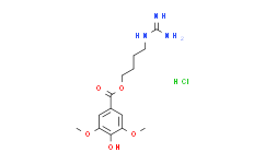
Leonurine hydrochloride is an alkaloid isolated from Herba leonuri, with anti-oxidative and anti-inflammatory. |
| In Vivo: |
Leonurine (10 mg/kg/d, p.o.) significantly increases the expressions of PPARγ, LXRα, ABCA1 and ABCG1, and decreases both TG and TC levels in serum of mice[1]. Leonurine hydrochloride (50, 100, 200 mg/kg) improves intracellular lipid accumulation via activating AMPK/SREBP1 pathway, enhances biochemical parameters, reduces hepatic lipoperoxide and increases antioxidant levels in mice[2]. Leonurine (20?mg/kg, p.o.) ameliorates osteoarthritis development in mouse DMM model[3]. |
| In Vitro: |
Leonurine (0, 5, 10, 20, 40, 80 μM) causes diminution in lipid accumulation, cellular cholesterol content, including total cholesterol (TC), free cholesterol (FC) and cholesteryl ester (CE), and increase in apoA-I- or HDL-mediated cholesterol efflux after treatment for 24 h. Leonurine also significantly and dose-dependently increases the expressions of ABCA1 and ABCG1 at the mRNA and protein levels in human THP-1 macrophages, and such effect is involved in PPARγ[1]. Leonurine hydrochloride (LH) shows protective effect on cell viability of HepG2 and HL-7702 cells incubated with palmitic acid (PA) of free fatty acid (FFA) for 24?h. Leonurine hydrochloride (125, 250, 500?μM) improves cellular lipid accumulation in HepG2 and HL-7702 cells via activating AMPK/SREBP1 pathway[2]. Leonurine (5, 10, 20?μM) inhibits the expression of iNOS, COX-2, PGE2, NO, TNF-α, and IL-6 in IL-1β-induced human chondrocytes, suppresses ECM degradation in human OA chondrocytes, and blocks IL-1β-induced PI3K and Akt phosphorylation in a dose-dependent manner[3]. |
| Cell Assay: |
MTT assay is performed to study the cytotoxic effects of Leonurinein HepG2 and HL-7702 cells. Briefly, HepG2 and HL-7702 cells are seeded for 24?h at the density of 3?×?104 cells/well in 96-well plates. After 24?h incubation, cells are treated with different concentrations of Leonurine (0-1000?μM) and the control group is treated with only DMEM for 24?h at 37°C in 5% CO2 incubator. Then, these cells are treated with MTT solution (5?mg/mL) for further 4?h. After 4?h incubation, DMEM containing MTT solution is discarded. Cells are then dissolved by adding DMSO (200?μL) to each well and the solutions are mixed thoroughly for 5?min. Finally, the absorbance is determined at 570?nm with a microplate reader[2]. |
| Animal Administration: |
MiceApoE-/- mice (male, eight-week old) are fed a chow diet for 2 weeks, apoE-/- mice are randomly divided into several groups (n=15/group). Mice in the Leonurine group are intragastrically administered with Leonurine (10 mg/kg/d) every day and continued for 8 weeks. The control group is fed with an equal volume of PBS. At week 16, mice are euthanized, followed by collecting the blood and tissue samples for further analyses[1]. |
| References: |
[1]. Jiang T, et al. Leonurine Prevents Atherosclerosis Via Promoting the Expression of ABCA1 and ABCG1 in a Pparγ/Lxrα Signaling Pathway-Dependent Manner. Cell Physiol Biochem. 2017;43(4):1703-1717.
[2]. Zhang L, et al. Novel hepatoprotective role of Leonurine hydrochloride against experimental non-alcoholic steatohepatitis mediated via AMPK/SREBP1 signaling pathway. Biomed Pharmacother. 2018 Dec 7;110:571-581.
[3]. Hu ZC, et al. Inhibition of PI3K/Akt/NF-κB signaling with leonurine for ameliorating the progression of osteoarthritis: In vitro and in vivo studies. J Cell Physiol. 2018 Nov 11. |


