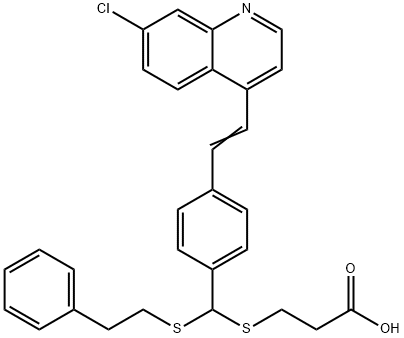
2449093-46-1
 2449093-46-1 結(jié)構(gòu)式
基本信息
物理化學(xué)性質(zhì)
常見問題列表
IC50: 24.5?μM (ATG4B); Kd: 16 μM (ATG4B)
LV-320 (0-120?μM; SKBR3, MCF7, JIMT1, and MDA-MB-231 cells) treatment results in a dose-dependent increase in endogenous LC3B-II and protein p62 levels in all four cell lines.
LV-320 (120?μM; 48 hours; MDA-MB-231 cells) treatment results in an increase in LC3B-II, indicating that LV-320 blocks autophagic flux.
Western Blot Analysis
| Cell Line: | SKBR3, MCF7, JIMT1, and MDA-MB-231 cells |
| Concentration: | 0?μM, 25?μM, 50?μM, 75?μM, 100?μM, or 120?μM |
| Incubation Time: | |
| Result: | Resulted in a dose-dependent increase in endogenous LC3B-II and protein p62 levels in all four cell lines. |
Cell Autophagy Assay
| Cell Line: | MDA-MB-231 cells |
| Concentration: | 120?μM |
| Incubation Time: | 48 hours |
| Result: | Blocked autophagic flux. |
LV-320 (100-200?mg/kg; oral gavage; three times over two days; GFP-LC3 mice) treatment results in a terminal blood level of 169 μM and a liver level of 104 μM. The expression of GFP-LC3 puncta is significantly greater accumulation in LV-320 treated animals compared to controls. LC3B-II protein is also increased in LV-320-treated animals. The treatment do not cause significant toxicity in mice at either dose.
| Animal Model: | GFP-LC3 mice (females, 9-14 weeks) |
| Dosage: | 100?mg/kg or 200?mg/kg |
| Administration: | Oral gavage; three times over two days (Pharmacokinetic study) |
| Result: | Terminal blood levels were 169?μM and liver levels were 104?μM. LC3B-II protein level was also increased. |